[Article] - TEM in situ crystallization of SmNiO3 nanoparticles
Abstract
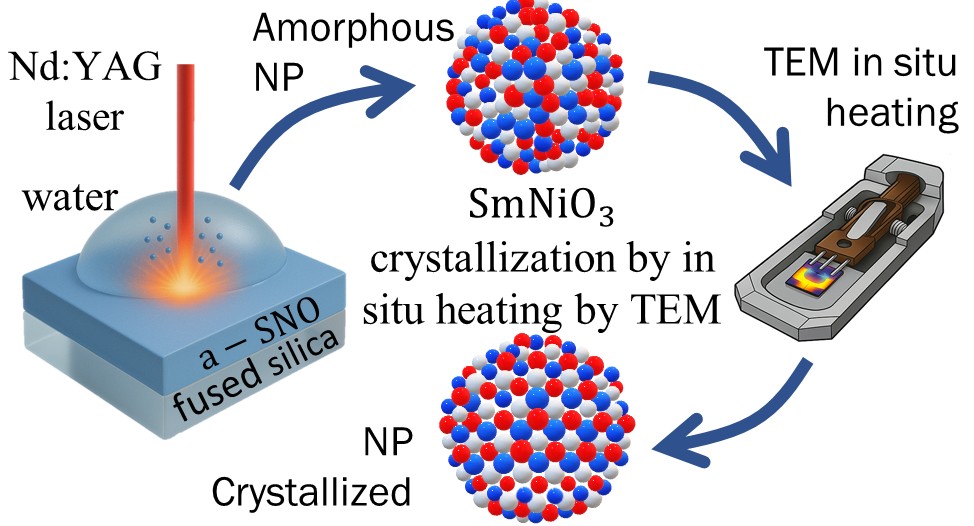
We report a direct in situ transmission electron microscopy (TEM) study of the crystallization of perovskite nickelate SmNiO3 from amorphous Sm−Ni−O nanoparticles. These nanoparticles, with a mean diameter of ∼30 nm, were generated by laser ablation of sputtered amorphous Sm−Ni−O films and deposited onto grids specially conceived to allow for in situ heating during TEM observations. In situ heating to 600 °C (ramp rate 600 °C/s, 30 s hold) within the TEM revealed the emergence of crystallized SmNiO3 domains, coexisting with secondary Sm2O3 or NiO phases in particles exhibiting off-stoichiometric Sm/Ni ratios. High-resolution TEM, fast Fourier transforms, and energy-dispersive X-ray spectroscopy demonstrated that initial chemical composition, rather than particle size, dictates phase evolution: near-stoichiometric particles form predominantly SmNiO3, whereas Sm- or Ni-rich particles undergo sequential crystallization of Sm2O3 or NiO followed by SmNiO3 nucleation. Core-loss electron energy-loss spectroscopy confirmed an order-of-magnitude increase in the Ni3+/Ni2+ ratio upon annealing, signifying perovskite formation alongside residual Ni2+ that can be attributed to oxygen vacancies or compositional inhomogeneities. A thermodynamic approach based on Gibbs free-energy landscapes rationalizes these pathways, and Gaussian fitting of SmNiO3 grain sizes versus Ni content reveals maximum domain growth at ideal 1:1 Sm/Ni stoichiometry. These findings elucidate nanoscale crystallization mechanisms in strongly correlated oxide nanoparticles and offer a new potential route to integrating this material in new functional device architectures.
Autors
Carlos Calvo-Mola, Stéphanie Bruyère, Vicente Torres-Costa, Silvère Barrat
References
J. Phys. Chem. C 2025, 129, 35, 15806–15814
DOI
https://doi.org/10.1021/acs.jpcc.5c04120